How Long Will Symptoms Of Brain Fog Last After Being Infected With Covid-19? How To Get Rid Of Long Covid-19 Brain Fog?
- Long Covid Classified Treatment Options
- 26 Oct, 2022
Summary
Numerous studies have confirmed that novel coronavirus infection in humans produced long-lasting side effects even after the symptoms of the initial infection disappeared completely, which we call long-term COVID-19 symptoms. These post-coronavirus remnants mainly were the appearance of brain fog, fatigue, shortness of breath, chronic pain, loss of smell, loss of taste, early symptoms Parkinsonism-like, early symptoms of Alzheimer 's-like disease and diarrhea. These long-term COVID-19 symptoms would put long-term adverse effects on people 's lives and work.
In this paper, the authors studied the long-term COVID-19 symptoms, finding that there was a large proportion of patients with the symptoms of brain fog. This article summarized the clinical manifestation of long COVID-19 syndromes of brain fog and gave an in-house self assessment to judge whether people developed these symptoms. Then, the pathogenesis of symptoms of brain fog caused by long COVID was analyzed, which included: the process of infection of human cells with COVID-19, virus replication in human cells to trigger pyroptosis, immune system response to the virus, virus transmission in the human body, virus invasion into the brain through the olfactory nerve, virus penetration into the brain through the blood-brain barrier, and virus causing neuroinflammation in the brain. Finally, combined with the pathogenesis, we gave the therapeutic ideas of western medicine for the disease after infection with COVID-19.
Due to the limitation of western medicine treatment against long COVID-19 symptoms of brain fog, this paper also recommended some traditional Chinese medicine, including Chinese medicine formula and acupuncture treatment, to treat the symptoms of brain fog. In addition, this paper made advises patients with long-term COVID-19 brain fog on taking healthcare products, as well as on exercise, diet, and sleep. In the end, we hope that the treatment program for long-term COVID-19 symptoms of brain fog suggested in this paper could benefit patients by improving their symptoms as soon as possible, and even cure these symptoms of brain fog caused by novel coronavirus.
Keywords: Long COVID, Post-COVID Conditions, long COVID brain fog, symptoms of brain fog after COVID-19 infection, brain fog induced by COVID-19, how to get rid of brain fog
1.What are The Clinical Manifestations of Long COVID Brain Fog?
A large proportion of long-term COVID-19 symptoms are associated with neurological symptoms, and some medical communities use a folk colloquial vocabulary” brain fog” to facilitate the unified classification and description of post-COVID-19 symptoms associated with the brain. Brain fog has not previously been regarded as a medical or scientific term, but rather a colloquial description of a state in which people cannot think normally and clearly when they develop influenza or other diseases. Ordinary people use the fog in their brains to describe problems arising in their usual cognition or thinking, such as dizziness, tinnitus, difficulty concentrating, memory loss, slower thinking speed, inability to think keenly without creativity, and difficulty formulating plans.
During rehabilitation after being infected with COVID-19, many patients said they experienced "brain fog". According to figures given in the study report, about 10% of patients infected with COVID-19 will experience long COVID. At present, it has become customary in the medical community to classify the symptoms of brain and nerve caused by COVID-19 infection as long-term COVID brain fog. According to the statistics in the study report, up to 60% of patients with long-term COVID presented some degree of brain fog.
According to the published medical papers on the symptoms of long COVID brain fog, we concluded clinical manifestations of brain fog as follows:
(1)Neurasthenia: feel that the brain is often in a state of confusion; without mentally lethargic; feel a slower thinking when pondering; respond slower when doing things; lose creativity when working; and feel neurasthenic.
(2)Memory loss: feel that one has a cognitive impairment; often unable to remember something; memory loss, forgetfulness; appear similar symptoms to senile dementia-like and Parkinson's disease.
(3)Attention disorder: feel that the brain is not in a state and mental trance; feel it is easy to distract and difficult to concentrate.
(4)Get lost: feel the brain often is in the state of vertigo; feel dull in discerning spatial directions.
(5)Language disorders: feel that it is not fluent and smooth when speaking; often forget to choose which words to express meaning.
(6)Dizziness: feel a false sense of movement or rotation, vertigo, feel lightheaded, unstable to stand or walk, and likely to lose balance and fall.
(7)Brain fatigue: feel drowsy, it’s easy for brain to feel tired, feel fatigued when thinking,it’s easy to feel sleepy when reading, feel sleepy in the daytime.
(8)Common accompanying symptoms,loss of smell: olfactory dysfunction, feel that one can not smell anything through the nose, or that the smell is abnormal.
(9)Common accompanying symptoms, blurry vision: blurred vision is presented in the morning, feel that the vision is in a trance and eyes are tired when staring at the computer plane and mobile phone screen for a longer time.
(10)Common accompanying symptoms,tinnitus: refers to the condition in which the external environment does not make a sound, but feels to hear a certain noise, feels that the ear is blocked by cotton, over-high internal pressure,buzzing, and it’s easy to be anxious or difficult to concentrate.
(11)Common accompanying symptoms,chronic headache: feel symmetrical chronic headache bilaterally on the head; often feel hot on the head and tightness on the head.
2.How Long Will Symptoms Of Brain Fog Last After Being Infected With Covid-19?
2.1 What Are Post-Covid Conditions?
According to the definition of the World Health Organization, Post-COVID Conditions or Long COVID, also known as Post COVID-19 syndrome, refers to since the patient was infected with the novel coronavirus, the results of his PCR showed positive as criteria, and he still suffers uncomfortable symptoms after 3 months. More detailed updated statistical information on the incidence of Post Covid-19 Symptoms according to the timeline can be found in the figure below.
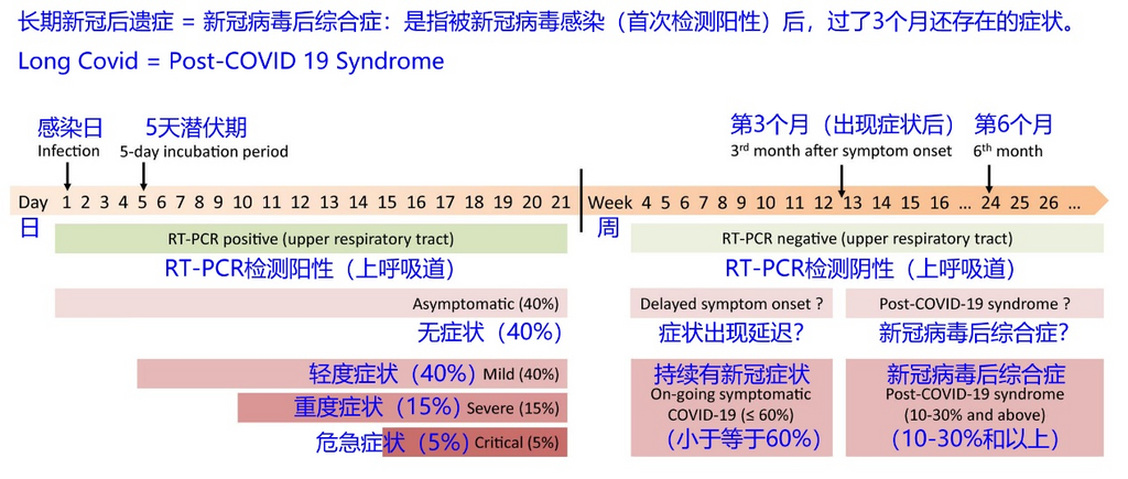
Post-COVID Conditions are mainly concentrated in five aspects, including respiratory syndrome, cognitive system syndrome, chronic fatigue syndrome, chronic pain syndrome, and mental syndrome, such as long-term COVID-19 brain fog, which belongs to the cognitive system syndrome. Long-term COVID chronic fatigue are one of chronic fatigue syndrome.
If you have a history of the novel coronavirus, which means your previous result of PCR test showed positive, and before infection with the novel coronavirus, you didn’t suffer from any brain fog symptoms, but now you are presenting these symptoms of brain fog in the above section, you are likely to afflict by Post Covid-19 Symptoms of brain fog.
2.2 How Long Will Symptoms Of Brain Fog Last After Being Infected With Covid-19?
In 2021, in a study designed to describe long COVID symptoms in more than 3,000 people from 56 countries, researchers found that 88% of respondents represented brain fog symptoms such as cognitive or memory problems. During the first few months after the onset of COVID-19, these symptoms showed an increasing trend and then started to decrease gradually. At the beginning of the 7th month after the onset of COVID-19, 55.5% of respondents reported suffering from cognitive problems such as brain fog.
In another study in 2021, patients with brain fog caused by long-term COVID were asked to describe their experiences. As in the previous study, most participants reported brain fog in the first few months following the onset of COVID-19. Participants were followed up by email for 4 to 6 months after the initial assessment. Of those who responded to follow-up, 65% people felt that their symptoms of brain fog were gradually improving.
A 2022 study investigated the rehabilitation of individuals with neurological symptoms such as long-term COVID and brain fog. The mean duration of brain fog since participants developed symptoms of COVID-19 infection was 14.8 months. After an initial evaluation, participants were followed up for 6 to 9 months. During follow-up, no significant changes were observed in the reporting of brain fog symptoms compared with the initial assessment, which means there were no significant change among most patients with brain fog. Researchers pointed out that quality of life measures of study participants remained lower than that of the general population.
According to published medical statistics papers on long COVID brain fog, the symptoms could last for quite a long time. The symptoms of brain fog tends to peak within a few months of infection with COVID-19 and them generally improve over time. Recent studies have found that brain fog symptoms may last more than a year after infection with COVID-19. Moreover, studies have shown that more than 20% of patients with long COVID brain fog still did not get an improvement for their symptoms after a year.
3.What Changes Our Bodies Will Experience After Being Infected With COVID-19?
The coronavirus infects the human body mainly through respiration, and the virus usually infects the nasal cavity, throat, and upper respiratory tract, then invades the epithelial cells of various organs and tissues of the respiratory system, and then into pericytes and blood. Finally, it spreads along the blood to various organs and tissues throughout the body. For long-term COVID brain fog is generally attributed to a state of brain inflammation which is directly or indirectly caused by novel coronavirus infection, according to published medical research papers.
According to the current medical research papers on brain fog symptoms after infection with COVID-19, the causes of brain fog can be broadly divided into the following causes:
(1) There is no COVID-19 in the human body, but the previous novel coronavirus infection has caused relatively large damage to various organs of the human body and the immune system of the human body. Proinflammatory cytokines and chemokines produced by these insults, as well as dead viral corpse fragments, trigger chronic inflammation in humans. Chronic inflammation in the human body leads to some pro-inflammatory factors breaking through the blood-brain barrier into the brain, which in turn triggers chronic inflammation in the brain.
(2) Trace amounts of COVID-19 remain in the human body, but they have been suppressed by the human immune system. Due to the minute amounts of viruses, it is hard for PCR nucleic acid to detect, thus the result presents a negative. These residual minute amounts of coronavirus, which still invade cells in various organs of the human body, trigger chronic inflammation in the human body. Chronic inflammation in the human body leads to some pro-inflammatory factors breaking through the blood-brain barrier into the brain, which in turn triggers chronic inflammation in the brain.
(3) A very small amount of novel coronavirus can also directly infect nerve cells. For example, the virus infect olfactory neuron cells directly through the nasal cavity into nasal epithelium, and then make an infection by Dynein protein, which is responsible for the retrograde transport of various cellular substances required from neuronal axons to the nucleus, and then by vesicles between the nerves in the brain to transmit to other nerve cells. This process can also trigger chronic inflammation of the brain.
(4) At the early stage of infection, the coronavirus enters the human brain directly through the olfactory organs, or breaks through the blood-brain barrier to infect the surrounding cells and microvascular endothelial cells in the brain, triggering chronic inflammation of the brain.
(5) There are a small number of novel coronavirus that can also directly infect the surrounding cells of neurons in the brain, such as horizontal basal cells and Sertoli cells. Damage to cells around neurons in the brain can decrease the function of neuronal cells, which in turn triggers various brain operation problems. This process can also trigger chronic inflammation of the brain.
3.1 The Process Of Covid-19 Infecting Cells And Finally Triggering Pyroptosis Through Cell Reproduction
The mechanism by which novel coronavirus infects human cells is that virions recognize host receptors through the coronavirus spike glycoprotein (S protein), enter host cells in a membrane-fused manner, replicate in host cells through large replicative transcription complexes, and promote proliferation by interfering with and inhibiting the host immune response. The hosts of human highly pathogenic coronavirus are humans and vertebrates, and virions infect lung cells and upper respiratory cells through droplets, contact, and aerosols, and may also infect and spread through other routes such as the digestive tract, urine, and eyes.
The following figure shows the process how the COVID-19 infect cells and reproduce through the cells, finally triggering pyroptosis.
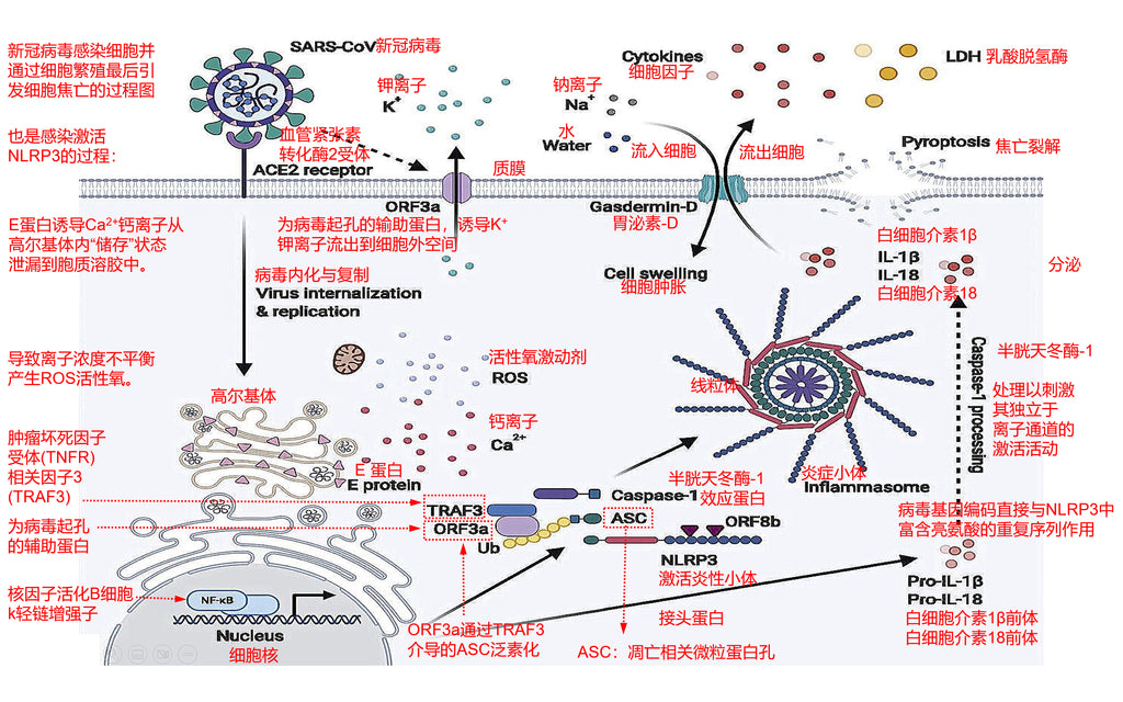
The approximate process is as follows:
(1) Novel coronavirus enters through the respiratory tract and attaches to epithelial cells of the upper respiratory tract and lungs.
(2) The novel coronavirus recognizes host receptors through the spike glycoprotein (S protein) of coronavirus, then binds to the angiotensin-converting enzyme 2 (ACE2) receptor and enters host cells in a membrane-fused manner.
(3)The novel coronavirus enters the Golgi apparatus and promotes the leakage of calcium (Ca2 +) ions stored in the Golgi apparatus by the E protein from the Golgi apparatus to the cytosol region.
(4) Simultaneous activation of ORF3a (a protein that opens pores in the cell membrane of the virus) , induces the efflux of potassium ions (K +) through the cytoplasmic membrane into the extracellular space.
(5) This imbalance in ion concentrations inside and outside the cell further leads to the generation of reactive oxygen species ROS oxidation reactions.
(6) In addition to inducing potassium ion (K +) efflux, ORF3a (a protein that opens pores in the cell membrane by the virus), also mediates ASC ubiquitination (protein ASC that drives apoptosis and open pores in the cell membrane) through TRAF3 (tumor necrosis factor receptor TNFR-related factor 3).
(7) This process in turn leads to mitochondrial damage and triggers activation of the NLRP3 inflammasome (tumor necrosis factor receptor TNFR-related factor 3).
(8) ORF8b directly interacts with leucine-rich repetitive DNA sequences of NLRP3 (tumor necrosis factor receptor TNFR-related factor 3) to stimulate its activation activity in independent of ion channels.
(9) NLRP3 inflammasome activates and induces the formation of gasdermin-D pores on the cell membrane, leading to IL-1 b and IL-18 secretion, which then spreads to the internal and external cellular environment.
(10) Water also flows into the interior of the cell through open holes in the cell membrane, which in turn leads to cell swelling and subsequent cell membrane rupture, which is pyroptosis.
(11) Novel coronavirus infection or vaccination can induce novel coronavirus specific antibodies (IgG) to adhere to novel coronavirus particles.
(12)Activated inflammasome NLRP3 (tumor necrosis factor receptor TNFR-related factor 3) in turn activates caspase-1 (caspase-1), resulting in gasdermin (GSDMD) cleavage.
(13)The cleaved NLRP3 inflammasome terminal N would be moved to the plasma membrane of the cell by GSDMD and assembled into pores in it.
(14) After the active replication of the coronavirus, the coronavirus would be released through emptiness on the cell membrane, or automatically after pyroptosis of the host cell.
3.2 The Process Of The Human Immune System Eliciting An Inflammatory Response In Order To Eliminate The Coronavirus
The response of the human immune system to the site of infection with novel coronavirus infection is divided into two phases: a preliminary phase in which the immune system mobilizes macrophages and polymorphonuclear phagocytes (e.g., neutrophils, basophils, eosinophils) to rapidly reach the site of infection to control infection. In the following stages: the immune system of the human body takes about 4 days to establish adaptive immune mechanisms through learning, including T cell-mediated immune mechanisms to solve the infection, and B system-mediated antibody production to clear the pathogen- novel coronavirus to prevent reinfection.
The following figure shows the process of the human immune system eliciting an inflammatory response in order to eliminate the novel coronavirus:
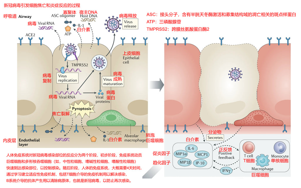
(1) Novel coronavirus enters through the respiratory tract and attaches to epithelial cells of the upper respiratory tract and lungs. The novel coronavirus recognizes host receptors via the spike glycoprotein (S protein) of coronavirus.
(2) Then the coronavirus binds to ACE2 (angiotensin-converting enzyme 2) receptor and TMPRSS2 (transmembrane serine protease 2) receptor to enter host cells in a membrane-fused manner.
(3) Novel coronavirus triggers DAMPS (Damage-associated molecular pattern) in cells leading to pyroptosis, after which cells release various factors, including ATP (adenosine triphosphate), nucleic acids, and ASC oligomers (adaptor molecules, apoptosis-associated speck-like protein with a caspase-recruitment domain).
(4) DAMPS (Damage-associated molecular pattern) is recognized by adjacent epithelial cells, endothelial cells, and alveolar macrophages. This in turn triggers the production of pro-inflammatory cytokines and chemokines including IL-6, IP-10, macrophage inflammatory protein 1α (MIP1α), MIP1β, and MCP1.
(5) These pro-inflammatory cytokine proteins attract monocytes, macrophages, and T cells produced by the immune system to the site of infection and then these cells begin to perform their duties to eliminate the novel coronavirus.
(6)At the same time, T cells will produce IFNγ in large amounts and establish a pro-inflammatory feedback loop. Pyroptosis also generates G-CSF (granulocyte colony-stimulating factor) and TNF (tumor necrosis factor).
(7) Immune cells accumulate further at the site of infection, resulting in excessive production of pro-inflammatory cytokines and further damage of the cellular environment at the site of infection, thus promoting a further inflammatory response.
There are trace viruses that escape in the human body in the long term, which will persistently infect and invade human tissue cells, causing tissue damage. Tissue damage, in turn, triggers chronic inflammation. This chronic inflammation is produced by a persistent immune response, also causing persistent massive cytokine diffusion. The continuous breakdown of a large number of cytokine proteins can lead to autoimmunity. Autoimmunity refers to the immune response produced by organisms against healthy cells and tissues of their bodies. Any disease caused by this immune abnormality is called autoimmune disease.
The persistence of trace novel coronavirus continues to deteriorate the situation, leading to T cell failure and immune memory defects. Defects in immune memory can make the immune system gradually insensitive to immune pathways that eliminate novel coronavirus, eventually becoming a coexisting state. This condition is characterized by generalized diffuse chronic inflammation. In the long run, the function of body organs and tissues is reduced.
Because novel coronavirus can easily mutate, so far as more and more coronavirus variants appear and the ability of immune escape is increasing, this makes the effect decrease when the antigens (HLA) produced by the human immune system to clear novel coronavirus, which means there will be a small number of novel coronavirus immune escape. Novel coronavirus in some patients has not yet been completely cleared, but because the human immune system is suppressing these coronaviruses, leading the content of novel coronavirus too small so as to make PCR tests negative.
3.3 The Process Of Novel Coronavirus Invading The Brain Through The Olfactory Nervous System And Leading To Brain Inflammation
In order to enter host cells, novel coronavirus uses the angiotensin-converting enzyme 2 (ACE2) receptor beside other substances. A study has shown that expressed could be observed in the central nervous system (CNS), more precisely, its expression could be observed in cerebral vessels, epithelial cells (CP) of a single layer of venation, and neocortical neurons. This expression pattern suggests that the virus may be able to enter the central nervous system.
Studies have shown that the coronavirus can trigger inflammation of the olfactory bulb or even atrophy of the olfactory bulb if it further invades epithelial cells in the olfactory bulb, such as capillary epithelial cells, which disrupts the transmission of electrical signals between olfactory neuron cells and mitral neuron cells. Based on the above studies, we can deduce the invasion process of novel coronavirus into the brain through the olfactory nervous system.
The following figure shows how the novel coronavirus invades the brain through the olfactory nervous system and leads to brain inflammation:
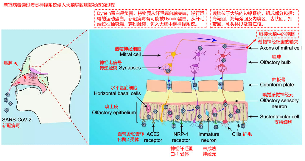
(1) Novel coronavirus enters through the respiratory tract and attaches to epithelial cells of the upper respiratory tract and lungs. The novel coronavirus recognizes host receptors via the spike glycoprotein (S protein) of coronavirus.
(2) It then binds to ACE2 (angiotensin-converting enzyme 2) receptor and TMPRSS2 (transmembrane serine protease 2) receptor and enters Sertoli cells of the olfactory epithelium and horizontal base cells of the olfactory epithelium in a membrane-fused manner.
(3) Extracellular vesicles (EV) are nanoscale membrane vesicles composed of lipid bilayers and secreted by all cell types. They act as carriers, protecting macromolecules such as proteins and RNAs from enzymatic degradation, and transporting these macromolecules between different cells, from adjacent cells to more distant cells, or immune cells. In addition, specific extracellular vesicles (EVs) have been shown to be able to cross the brain barrier.
(4) Very small amounts of novel coronavirus in Sertoli cells of the olfactory epithelium and horizontal base cells of the olfactory epithelium may be encapsulated into extracellular vesicles (EVs) and then enter olfactory neuronal cells.
(5) In neuronal cells, Dynein protein is the motor protein responsible for retrograde transport of substances from the cilia end to the axons end. The novel coronavirus is likely to be pulled from the cilia end of olfactory neurons to the axonal end by the Dynein protein, thereby entering the olfactory bulb.
(6) In the olfactory bulb, olfactory neurons and mitral neurons communicate neuroelectrical signals through touch process connections, at which time novel coronavirus may be encapsulated into extracellular vesicles (EVs) and then into mitral neuron cells.
(7) In mitral neurons, novel coronavirus may be retrogradely transported by Dynein protein into deeper brain neurons and thus enter the central nervous system of the brain.
The brain region connecting the olfactory bulb is called the limbic system of the brain, also known as the olfactory brain. If the virus further invades the olfactory brain, which is the part of the limbic system of the brain that is responsible for the processing of logic operations for the encoding and decoding of olfactory electrical signals, this makes the electrical signals of the olfactory nervous system unable to be normally understood and operated by the brain, which is the cause of cognitive dysfunction and also one of the causes of brain fog symptoms.
3.4 The Process Of Novel Coronavirus Invading The Brain Through The Blood-Brain Barrier Or The Parenchyma Of Ventricular Organs And Resulting In Brain Inflammation
According to the current medical research results and the generally agreed consensus, there may be the following four ways for the novel coronavirus to directly invade the brain:
(1) The coronavirus invades the brain directly through olfactory organ tissues and the olfactory nervous system.
(2) The coronavirus crosses the blood vessels, breaks through the blood-brain barrier, and directly invades the brain.
(3) The coronavirus passes through blood vessels and leaks to the brain at certain weak spots in ventricular organs, where these endothelial cells are not tightly connected and also lack an astrocyte interception barrier.
(4) The coronavirus infects peripheral nerve cells in human organ tissues, and is transported retrogradely to the brain end through the Dynein protein, and then leaks to the brain by latency into extracellular vesicles (EV). This possibility has not yet been fully substantiated.
Section 3.3 of this article explains how coronavirus invades the brain through olfactory organ tissues and the olfactory nervous system. The following article then explains the process of the coronavirus invading the brain through the blood-brain barrier or the parenchyma of ventricular organs, as shown in the following figure:
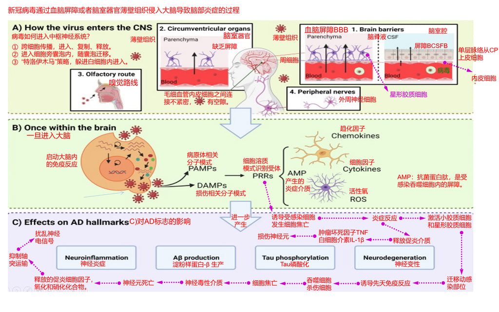
(1) The blood-brain barrier is usually composed of two modes: one is composed of an intercepting network combined of dense astrocytes, and the other is composed of a dense monolayer choroid plexus (CP) epithelial cells to form the blood-brain barrier BCSFB..
(2) The coronavirus is transported to the central nervous system through capillary blood, then infects endothelial cells of microvessels and crosses them, and then breaks through the intercepted network composed of dense astrocytes to enter the brain ventricular lumen.
(3) The coronavirus is transported to the central nervous system via capillary blood and then infects the endothelial cells of the microvessels and crosses them, then infects the monolayer choroid plexus (CP) epithelial cells and crosses them to enter the brain ventricular lumen.
(4) In addition to the blood-brain barrier, novel coronavirus enters the brain ventricular lumen from these loopholes in certain weak spots of ventricular organs, where these endothelial cells are not tightly connected and there is also a lack of astrocyte interception barrier.
(5) Once the coronavirus enters the brain, it begins to invade tissues and organs and central neuronal cells in the ventricular cavity, and start to replicat, proliferation, propagation.
(6) Viral infection activates the immune system in the brain and turns on PAMPs (pathogen-associated molecular patterns) and DAMPs (damage-associated molecular patterns), which in turn produce PRRs (cytosolic pattern recognition receptors).
(7) PRRs (cytosolic pattern recognition receptors) include: Cytokines (cytokines), Chemokines (chemokines), ROS (reactive oxygen species), AMP (antimicrobial protein peptides, which are inflammatory mediators and barrier fragments in infected phagocytes).
(8) At this point, the inflammation of the brain begins to spread.
From the above pathological deduction steps, novel coronavirus infection triggered an inflammatory response in the brain, which in turn produced a series of neurodegenerative lesions, causing long-term COVID-19 symptoms and being the cause of brain fog symptoms. When we develop long COVID symptoms of brain fog, we must treat it seriously, for brain fog will further develop into early Alzheimer's disease or Parkinson's disease. Therefore, medical attention should be sought as soon as possible when symptoms of brain fog occur.
4.What Are The Pathological Causes Of Brain Fog Symptoms After Infection With Covid-19?
4.1 Effects Of Sars-Cov-2 Infection On Neuroinflammation
Published studies have reported that 36.4% of SARS-CoV-2 inpatients presented with neurological symptoms, such as anosmia, headache, disturbance of consciousness, seizures, and stroke. In addition, a study in rhesus monkeys supported the ability of SARS-CoV-2 to enter the central nervous system. After intranasal inoculation of these non-human primates, the virus is able to enter the central nervous system, mainly through the olfactory bulb and spread to functional areas such as the hippocampus. In addition, infection is accompanied by pathological damage such as inflammatory response and neurodegeneration of the central nervous system.
However, since SARS-CoV-2 protein was detected only in very few cells, no viral particles were found in the brains of these rhesus monkeys and no effective replication in vitro appeared to occur in cell lines associated with the central nervous system (CNS). The researchers concluded that SARS-CoV-2 inside the CNS could not effectively infect and replicate, pathological damage is caused by cytokines in the central nervous system or systemic inflammation. The fact that SARS-CoV-2 replication and infection was inefficient in the central nervous system (CNS) may also explain why controversial results were obtained when SARS-CoV-2 was detected in the brain and cerebrospinal fluid of postmortem COVID-19 patients, that is, viral gene fragments were sometimes detected and viral gene fragments were sometimes undetectable.
Inflammatory cell infiltration was detected perivascularly in the hippocampus and medulla oblongata, suggesting SARS-CoV-2 infection induces neuroinflammation. Observed neuroinflammation can be caused by systemic inflammation and/or direct viral invasion of the brain. Yang et al. investigated postmortem brains of COVID-19 patients and observed that genes involved in inflammation (e.g., interferon) and complement pathways were upregulated in a monolayer choroid plexus epithelial cells.
In addition, monolayer choroid plexus epithelial cells signals from the epithelium of epithelial cells (CP) to the cortex. This communication is associated with the complement pathway that signals microglia and the inflammatory pathway that signals glia and neurons. As a result, microglia and astrocytes are activated and contribute to neuroinflammation. Yang et al looked more closely at subsets of microglia and astrocytes in postmortem brains, the gene expression profile of COVID-19-associated microglial subpopulations was observed to overlap with Alzheimer 's disease (AD) -associated microglia, whereas COVID-19-associated astrocyte subpopulations were mainly characterized by increased expression of glial fibrillary acidic protein (GFAP) and inflammatory factors as IFITM3 (interferon-induced transmembrane protein 3, antiviral effect).
In addition, the magnitude of the induced inflammatory response may depend on the genetic background of the patient. Magusali et al. showed that single nucleotide polymorphisms in the OAS1 gene increased the risk of Alzheimer 's disease (AD) development, and that associated variants of this gene were also associated with the severity of COVID-19. The OAS1 gene was expressed in microglia and appeared to be involved in limiting pro-inflammatory responses. Because OAS1 variants were expressed to a lesser extent than their wild-type counterparts, patients carrying OAS1 variants may develop more inflammation, which may make these SARS-CoV-2 infected patients more susceptible to Alzheimer 's disease (AD).
4.2 Effects Of Sars-Cov-2 Infection On Amyloid-β (Aβ) Accumulation
Yang et al reported increased IFITM3 (interferon-induced transmembrane protein 3, antiviral effect) expression in astrocytes and neurons of SARS-CoV-2 infected individuals, and IFITM3 showed increased amyloid-β40 (Aβ40) and amyloid-β42 levels, indicating that the latter also occurs with SARS-CoV-2 infection. The decreased clearance of amyloid-beta (Aβ) clearance may also be due to failure of amyloid-beta (Aβ) -destroying microglia in response to systemic inflammation.
In addition, the interaction between amyloid-β42 and the S protein of SARS-CoV-2 may decrease amyloid-β42 clearance. The latter was demonstrated by Hur using C57BL/6 mice. More precisely, deposition of amyloid-β42 in the blood of these mice was observed when amyloid-β42 was injected intravenously along with the extracellular domain of S protein. In summary, increased amyloid-β (Aβ) production and decreased clearance ultimately lead to increased amyloid-β (Aβ) deposition.
Increased levels of amyloid-β40 (Aβ40) and amyloid-β42 in neuronal extracellular vesicles (EV) were detected in both COVID-19 patients with and without neurological symptoms, further supporting this contention.
4.3 Effects Of Sars-Cov-2 Infection On Tau Phosphorylation
Transgenic Alzheimer 's disease (AD) mice showed increased tau pathology following injection of hepatitis virus (MHV) in a human coronavirus model mouse. In addition, hyperphosphorylation of Tau protein was observed in 3D scanned human brain organoids following SARS-CoV-2 infection. Moreover, elevated levels of phosphorylated tau protein were detected in neuron-derived extracellular vesicles (EV) in COVID-19 patients with and without neurological symptoms. This induction of Tau phosphorylation may be caused by pro-inflammatory stimulation of kinases of phosphorylate Tau.
4.4 Effects Of Sars-Cov-2 Infection On Neurodegeneration
Finally, neuroinflammation, amyloid-beta (Aβ) accumulation, and Tau phosphorylation can lead to neurodegeneration, a process that may also occur in COVID-19 patients. The presence of neurodegeneration was supported by elevated levels of neurofilament (NFL) peptides in cerebrospinal fluid, plasma, and neuronal extracellular vesicles (EV) in COVID-19 patients compared to controls, as well as neurodegeneration observed when SARS-CoV was intranasally inoculated into rhesus monkeys. Coronavirus itself and associated pyroptosis-induced NLRP3 activation could also support the neurodegeneration process.
The combination of the aforementioned Alzheimer's disease (AD) features with synaptic loss and decreased expression of neurotransmittance-related genes in excitatory neurons (also reported in COVID-19 patients) may lead to persistent clinical Alzheimer' s disease (AD) symptoms. Studies have shown that COVID-19 and Alzheimer's disease (AD) promote each other, and Alzheimer's disease (AD) patients may also be more likely to experience severe COVID-19 symptoms. A sign of this claim is that mortality and vulnerability to COVID-19 appear to be higher among people with dementia.
Amyloid-β42 has also been shown to bind to the S1 spike protein of SARS-CoV-2 and the human angiotensin converting enzyme 2 (hACE2) receptor conjugated to human angiotensin converting enzyme 2-Fc (hACE2-Fc). In addition, when immobilized S1 spike protein was first incubated with amyloid-β42 and then with hACE2-Fc, the binding strength of S1 spike protein to hACE2 (human angiotensin converting enzyme 2) increased.
In summary, there has been sufficient evidence that novel coronavirus infection causes chronic inflammation of the brain, which in turn leads to brain fog symptoms. Moreover, brain fog can cause the production of neurodegenerative markers, which means brain fog has the potential to develop into early Alzheimer's or Parkinson's disease.
5.How To Treat The Long Covid Brain Fog?
At first, we need to take a thorough physical examination of our bodies, and besides taking routine examinations, we need to do another four examinations.
The first item: do several more PCR tests, nasal and throat secretions should be dipped enough to check whether the result shows positive. If it shows positive, it means there is still some active novel coronavirus in the body.
The second item: perform a neurological examination. Because neurological symptoms are highly prevalent in patients with long-term COVID, neurological testing is recommended in all patients with long COVID-19, even if some patients communicate with their physicians, they do not have neurologically uncomfortable feedback. Many patients will report "brain fog", "difficulty in concentrating" and "poor memory." Physicians should carefully evaluate the patient 's subjective symptoms on a case-by-case basis to design a patient' s neurological examination to determine pathophysiology.
Following unconventional deeper neurological tests can be performed. For example, to rule out testicular brain injury, physicians are advised to perform functional magnetic resonance imaging MRI of the brain, including functional magnetic resonance imaging MR angiography of the brain. In elderly patients, imaging findings of early abnormal brain changes in Parkinson 's disease, Alzheimer' s disease, or other types of neurodegenerative dementia can be excluded by functional magnetic resonance imaging MRI of the brain.
When patients report memory impairment, it is important to determine whether this memory impairment is caused by infection with COVID-19 or whether COVID-19 infection worsens pre-existing symptoms. If ischemic or hemorrhagic stroke is observed on functional magnetic resonance imaging MRI of brain, we must consider medical therapy to prevent recurrent stroke.
Perfusion imaging (single photon emission computed tomography) may also be performed if the patient is wealthy or if the physician suspects the diagnosis. Cerebral perfusion imaging, which is perfusion imaging using CTP and MRP, has become a routine method to examine cerebral blood flow perfusion in stroke patients. Although there is still no evidence that perfusion imaging is an essential test for stroke assessment, many centers have begun to use perfusion imaging to assess cerebral blood flow in patients.
Cerebral perfusion testing may also show the inflammation triggered by non-specific immunity (innate immunity, which refers to the normal physiological defense function congenitally possessed by the body and can make the corresponding immune response to the invasion of various pathogenic microorganisms and foreign bodies) , which is characterized by hypoperfusion in the prefrontal or temporal lobes. Although there is no established treatment for this problem, some drugs that develop brain blood may help improve the condition.
Third item: in addition to basic blood tests, we assessed thyroid hormones, zinc, ferritin, antinuclear antibodies, rheumatoid factor, and blood sedimentation rate in almost all patients. Patients with PASC may have electrolyte disturbances, anemia, thrombocytopenia, hypoalbuminemia, lipid abnormalities, and abnormal glucose metabolism. These data need to be combined with patient symptom descriptions to carefully analyze the pathogenesis behind reasoning to determine whether abnormal laboratory test results can explain the symptoms of PASC.
The fourth item: listen to the sounds of the heart and respiratory system with a stethoscope. Anemia, the signs of heart failure, latent arrhythmias such as atrial fibrillation, and pneumonitis from enteritis must be assessed when abnormal sounds are detected, or if breathlessness persists, and to check whether oxygen saturation decreases during exercise.
The following items, from item 5 to item 7, are required when the patient presents other symptoms.
Fifth item: rheumatoid arthritis or other related diseases must be analyzed when patients have symptoms of joint discomfort.
Sixth item: when the patient complains hair loss, we examine the scalp, return to the history of hair loss symptoms, and confirm the weekly amount of hair loss.
Seventh item: neuropsychological tests should also be performed if the patient has significant psychological problems.
Then, the results of the above comprehensive physical examination and four special examinations should be handed over to the doctor, and then the person should communicate with the doctor to describe one’s own long COVID brain fog symptoms in detail.
Additionally, the doctor can design the best treatment for you. Because the situation varied from person to person, after the interview, the treatment plan given by the doctor is also different, so this paper cannot give a general treatment plan.
The rough ideas for designing the treatment plan are as follows:
(1) According to the results of the patient's comprehensive physical examination and four special examinations as well as the communication with the patient, find out the most likely pathogenesis of the patient's long-term COVID-19 symptoms of brain fog, namely exploring the reason.
(2) Prescriptions are prescribed for treatment according to the pathogenesis and the patient 's physical condition. In most cases, the brain is found to be slightly inflamed, and at this time, treatments against neuroinflammation are usually used.
(3) For the main direction of medication, if novel coronavirus remains in the body, antiviral drugs such as Paxlovid should be used as a priority. If there is no virus in the body, the drug is used from the perspective of reducing or eliminating the chronic inflammation of the patient, improving the metabolic circulation and immune mystery of patients. For example, rintatolimod (immunomodulator) and coenzyme Q10 + NADH (mitochondrial modulator) can be used to improve the immune system of patients.
(4) If Alzheimer 's disease are diagnosed, Lecanemab is needed to control the development of the disease. Eisai and Biogen announced that Lecanemab, an Alzheimer 's disease (AD) drug jointly developed by the two sides, has achieved good results in clinical trials for the treatment of patients with mild Alzheimer' s disease and Alzheimer 's disease, resulting in mild cognitive impairment (MCI).
(5) In case of psychiatric problems, a psychologist is needed. Massage of the brain can also be tried, and studies have shown that this is also beneficial for recovery.
6.How To Treat Long Covid Brain Fog From a Traditional Chinese Medicine(Tcm) Perspective?
From the theory of syndrome differentiation and treatment of TCM, the treatment of long-term COVID brain fog obtain more accurate TCM syndrome differentiation and more suitable TCM formulas from the following three steps.
(1) Perform the observation, smelling, hearing, and inquiring of traditional Chinese medicine diagnosis, and consult the comprehensive physical examination report and four special examination reports of western medicine in the previous section at the same time. Only in this way can TCM doctors obtain the most detailed disease information of patients to support them to make the most accurate syndrome differentiation and treatment of patients' conditions.
(2) TCM doctors will classify and summarize the main symptoms, accompanying secondary symptoms, physical condition, and sick parts of the internal organs of the patients according to the most comprehensive disease information of the patients. First, the main symptoms are the most unbearable symptoms of patients at present, such as dizziness. Second, accompanying secondary symptoms are that suffered from the main symptoms, which means the patient is experiencing other uncomfortable symptoms, such as occasional chronic headache. Third, to find out the physical condition of the patient, which means the patient's current physical health status, belongs to which category in the TCM constitution. For example, yang-deficiency constitution, phlegm-dampness constitution, qi-deficiency constitution, blood stasis constitution, etc.
(3) TCM doctors, according to the above summary, combined with the internal organs and meridians related to symptoms, comprehensively use the theory and practical experience of TCM to make TCM prescriptions for patients. If acupuncture and moxibustion are required, these programs will also be prescribed.
Traditional Chinese medicine (TCM) theory is mainly based on: Zhang Zhongjing’s “Treatise on Febrile Diseases”, “Synopsis of Golden Chamber”, Huang Yuanyu's “Four Sacred Hearts Source”, “Typhoid Fever Suspension”, “Jinkui Suspension”, “Changsha Yao Jie”, Li Dongyuan 's “Treatise on the Spleen & Stomach”, and Zhang Jingyue' s “Jingyue Quanshu”.
TCM Practical experience, which can be found in Google Scholar (Fig. https://scholar.google.com/), and search for keywords for TCM treatment of long-term novel coronavirus symptoms. For example, COVID-19 Traditional Chinese Medicine, Long Covid Traditional Chinese Medicine. A large number of academic papers on medical research on the treatment of Long Covid by traditional Chinese medicine can be found in this way.
In order to make it easier for everyone to understand the treatment process of TCM, the following is a TCM treatment case of a patient with long-term COVID-19 symptoms of brain fog.
Case 1:
Name: Li, male, aged 45 years, height 168cm, weight 90 kg.
Medical History: PCR was positive for novel coronavirus on 14 Mar 2022, followed by typical symptoms of novel coronavirus infection: fever, cough, sore throat, sputum with blood streaks, ageusia, and anosmia. Two weeks later, the symptoms of novel coronavirus infection gradually relieved and disappeared. Later, symptoms of brain fog appeared in succession.
Physical examination report of western medicine: (1) PCR test for novel coronavirus showed positive results again. (2) Cardiac examination revealed irregular heartbeat. (3) Proinflammatory markers were high and there was slight systemic chronic inflammation. (4) Immune system examination items revealed abnormal lymphocyte subset determination, C-reactive protein, and immunoglobulin data.
Main symptoms: brain fog.
Accompanying secondary symptoms: occasional chronic headache.
Prescriptions given by Western physicians:
(1) Paxlovid (antiviral drug, use to prevent the replication of novel coronavirus in vivo).
(2) Coenzyme Q10 + NADH (mitochondrial modulator).
(3) rintatolimod (immunomodulator).
(4)Lecanemab, a new drug jointly developed by Eisai and Biogen, controls the development of the disease.
(5) Vitamin C, Vitamin D, Vitamin E, Sulforaphane and resveratrol. (Use to reduce the inflammatory reaction in vivo.)
TCM constitution: the body presents phlegm-dampness constitution.
TCM diagnosis evaluation: exogenous wind evil, Xifeng Tongluo, resulting in dizziness symptoms.
Prescription given by the TCM doctor:
(1) Banxia Tianma Pills are used to invigorate the spleen and removing dampness, reducing phlegm and wind, and lessen dizziness symptoms.
(2) Mailuotong capsule is used to dredge meridian vascular congestion.
(3) Xifeng Tongluo Headache Tablets are used to clear away heat and toxic substances and reduce the symptoms of chronic headache.
Acupuncture program given by the TCM: electricity is exerted to heat the moxibustion apparatus, and apply moxibustion at the following acupoints.
(1) Baihui, Ashi, Shenyu and Guanyuan points to improve brain metabolism.
7.Which Kind Of Health Products Are Beneficial To Improving Long Covid Brain Fog?
Taking the patient above as an example, we give the following health product recommendations based on her condition:
(1) Omega 3 (Ω3) fatty acids, containing high amounts of DHA and EPA, are used to improve brain function and protect against cardiovascular and cerebrovascular diseases.
(2) VC, VD and VE are used to scavenge free radicals, anti-oxidation, and reduce systemic chronic inflammation.
(3) Curcumin is used to scavenge free radicals, anti-oxidation, activate NRF2, and reduce systemic chronic inflammation.
(4) Sulforaphane is used to scavenge free radicals, anti-oxidation, activate NRF2, and reduce systemic chronic inflammation.
(5) American ginseng is sliced for making tea or taken American ginseng capsules to replenish qi.
8.What Exercises Are Beneficial To Improving Long Covid Brain Fog?
Taking the patient above as an example, we give the following exercise advice based on his condition:
(1)Do mindfulness meditation or Buddhist meditation.
(2)Enjoy the wild of natural oxygen bars, such as walking in the deep forest, and perform deep breathing exercises.
(3)Assist with head massage.
9.What Is Beneficial To Improving Long-Term Covid Brain Fog In Terms Of Diet And Sleep?
Taking the patient above as an example, we give the following diet and sleep advice based on his condition:
(1) Control carbohydrate intake, which means reducing the amount of staple food such as rice and noodle, and control animal fat intake.
(2) Do not take any food after lunch, and have breakfast and lunch only a day. Losing weight at least more than 10% of body weight, which means losing 9 kg, reducing the degree of obesity.
(3) Bathing until slightly sweating before sleeping, and lie in bed to do general muscle tightness and relaxation training, improving sleep quality.
10.Patients With Brain Fog After Infection With Covid-19 Are Welcome To Contact With Our Long Covid Care Center
If there are similar symptoms among readers, contact our Long COVID Care Center for assistance.
Phone: +852 5765 5768
Whatsapp: +852 5765 5768
WeChat: longcovidcarecenter
Email: support@longcovidcarecenter.org
【Disclaimer: The treatment of diseases is a very complex and professional affair. Due to the limitation, Long COVID Care Center can only carry out remote simple interviews, unable to face-to-face offline interviews and obtain comprehensive physical examination results. Therefore, the suggestions, guidance, protocols, and documents conveyed by Long COVID Care Center to patients can only be used as a reference for patients to understand their diseases in many aspects, but cannot be directly used as a treatment plan. Patients must discuss their symptoms with doctors in local hospitals through face-to-face communication. After the patients completed the physical examination required by doctors, they would get a prescription issued by doctors and get the treatment under the guidance of doctors. Therefore, Long COVID Care Center hereby declares that our center is completely exempted from liability when any adverse consequences are caused by self-treatment of the patient for applying any contents convoyed by the center, that is, we do not bear any responsibilities.】


